
Guest post by Tamara A. Fulop, MD, Director, Breast Imaging at Mount Sinai Beth Israel
 Winning the battle against breast cancer is becoming a reality for women of all ages, thanks to diagnostic technology that can frequently detect breast cancer in its earliest stages. “Digital mammography acquires images electronically, resulting in better contrast, so we can often pick up smaller abnormalities before they become cancerous,” says Tamara A. Fulop, MD, Director of Breast Imaging at the Appel-Venet Comprehensive Breast Service at Mount Sinai Beth Israel.
A better picture
The combination of a well-performed breast examination and breast imaging can often detect breast cancer in its earliest stage – before a woman can detect a lump during a breast self-exam.
Mammograms are still the gold standard for screening in the early detection of breast cancer. In fact, an estimated 80 percent of breast cancers are found using screening mammograms. All women should get a baseline mammogram at age 40 and every year thereafter if the results are normal. Women with a family history of breast or ovarian cancer could require earlier mammographic screening and should speak with their doctors.
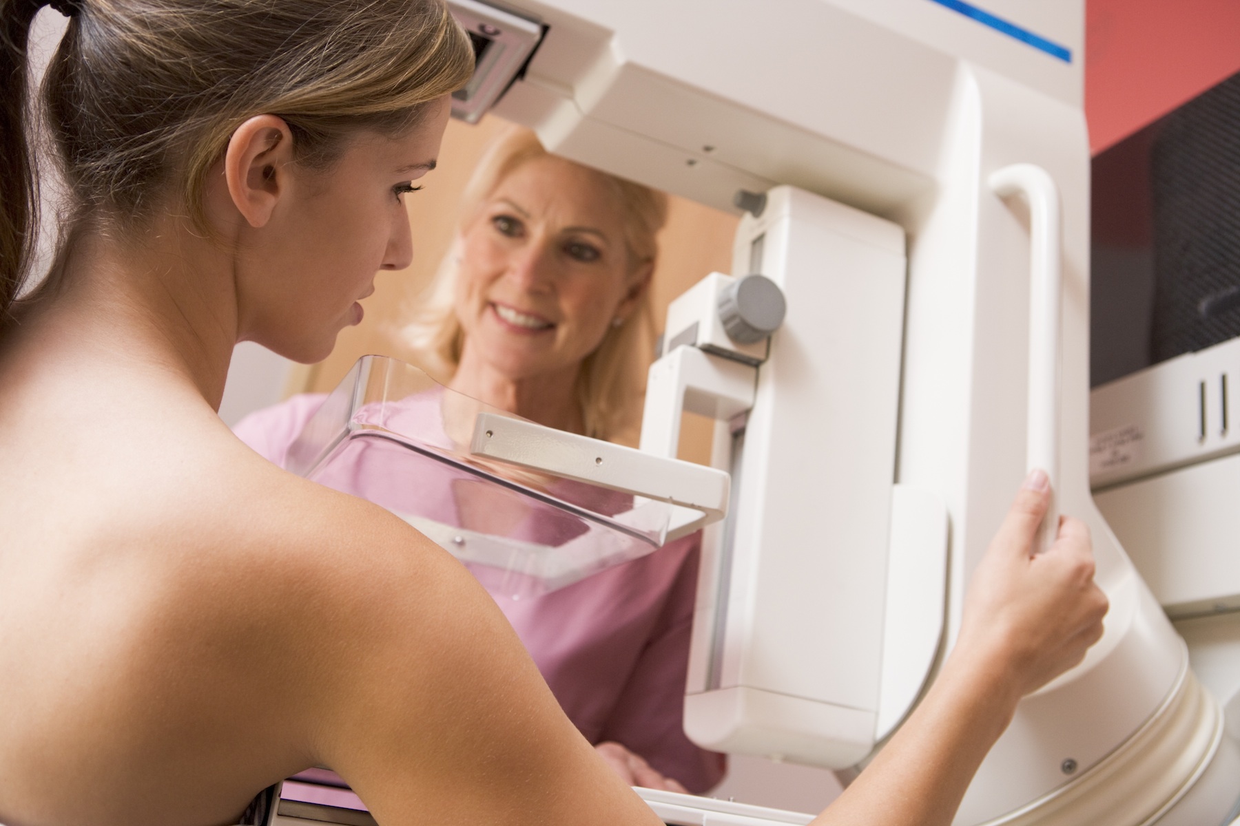
Digital mammograms have the potential to detect breast cancer at an earlier stage than traditional X-ray mammograms, and early detection is a key factor in breast cancer survival. Like conventional mammography, digital technology creates top-to-bottom and side-to-side images of each breast, but with a lower dose of radiation.
With digital mammography, images are available for viewing in seconds. “It can pick up calcifications and smaller nodules in patients younger than age 50, and in women who have dense breast tissue,” Dr. Fulop explains.
Expertise matters
The Breast Center at Mount Sinai Beth Israel also offers additional diagnostic services in one location:
Breast MRI is useful for high-risk patients, such as those recently diagnosed with a new breast cancer, who have a first-degree relative with breast or ovarian cancer, who are BRCA 1 or 2 gene positive, or who are of Ashkenazi Jewish heritage. A breast MRI creates multiple cross-sectional images of the breast using magnetic fields and a contrast agent.
Breast Ultrasound can pinpoint the location, size and shape of a lump and is used to evaluate certain kinds of abnormalities seen on mammogram. It can also be used to confirm whether a mass is solid or cystic. Ultrasound can also be utilized for the performance of image-guided needle biopsies.
Breast-Specific Gamma Imaging is a nuclear medicine exam, which can help identify cancerous breast tissue. This exam is similar to breast MRI but is useful if a patient is claustrophobic or larger-breasted.
“Technology is great, but it’s even more important to look for expertise and experience at the facility where you choose to have your breast imaging performed,” Dr. Fulop says. “If we find an abnormality on a routine mammogram, we bring the patient back for additional imaging. We have excellent communication with our referring physicians and a referral system in place so that patients can be cared for fairly seamlessly.